Diagnostic medicine is where disease becomes something you can see. A tissue section, a culture plate, a variant call. I am drawn to that moment of evidence and to the responsibility of reading it correctly.
Diagnostic medicine
The diagnosis that every other specialty depends on
In the clinic I sent samples away and waited. Now I work on the other side of that wait. My postdoctoral research at Hokkaido University uses whole slide imaging and spatial biology to ask what the tissue around a cell says about its state, and whether a computational prediction survives contact with morphology.
I am equally drawn to the molecular and microbiological sides of the field. A pathogenic BRCA2 variant, a resistant Helicobacter pylori isolate, and a T cell dysfunction score on a slide are all the same kind of object to me: evidence a clinician can act on.
Three kinds of evidence
Tissue, molecule, microbe
Anatomic and digital pathology
Whole slide imaging and spatial biology
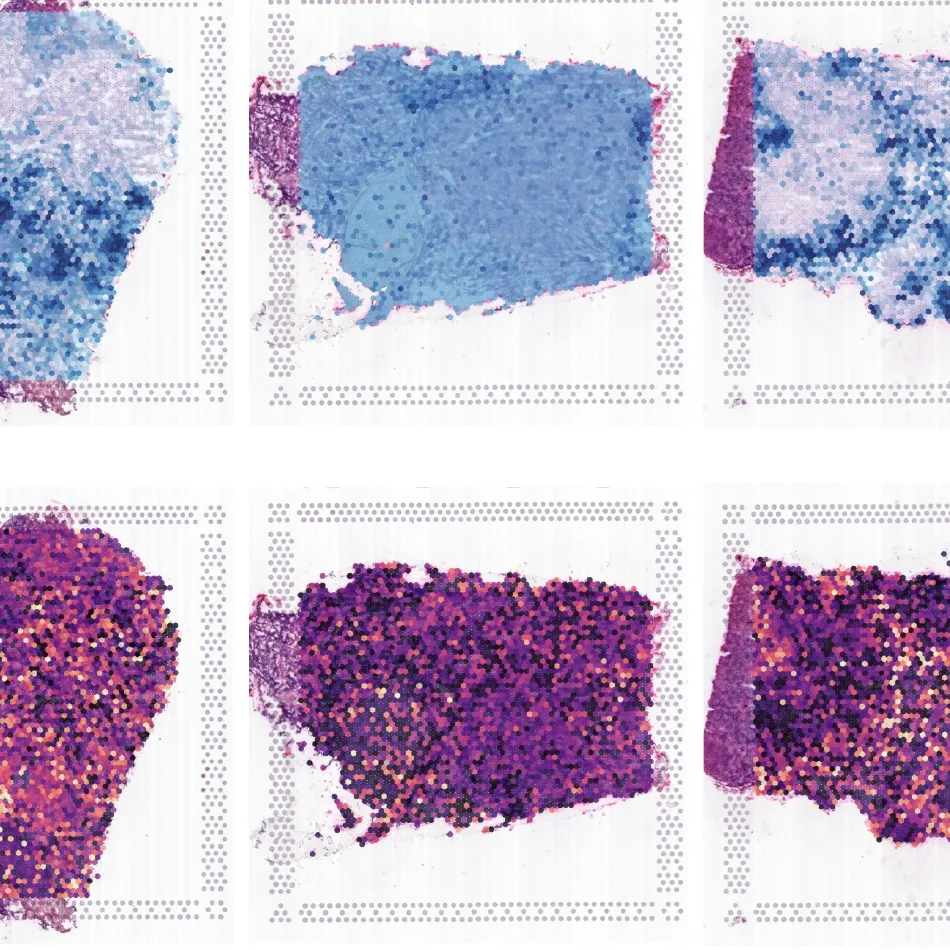
I analyse whole slide images alongside spatial molecular measurements. The question is always whether a predicted cell state makes sense in the tissue around it. A model can propose. The slide decides.
Molecular pathology
Variants that change management
I co-authored work on a pathogenic BRCA2 splice variant in metastatic prostate cancer and on the EHBP1 rs721048 variant in prostate and colorectal cancer in the Kazakh population. Both papers ask how a genomic finding should reach the patient.
Clinical microbiology
Culture, resistance, and surveillance
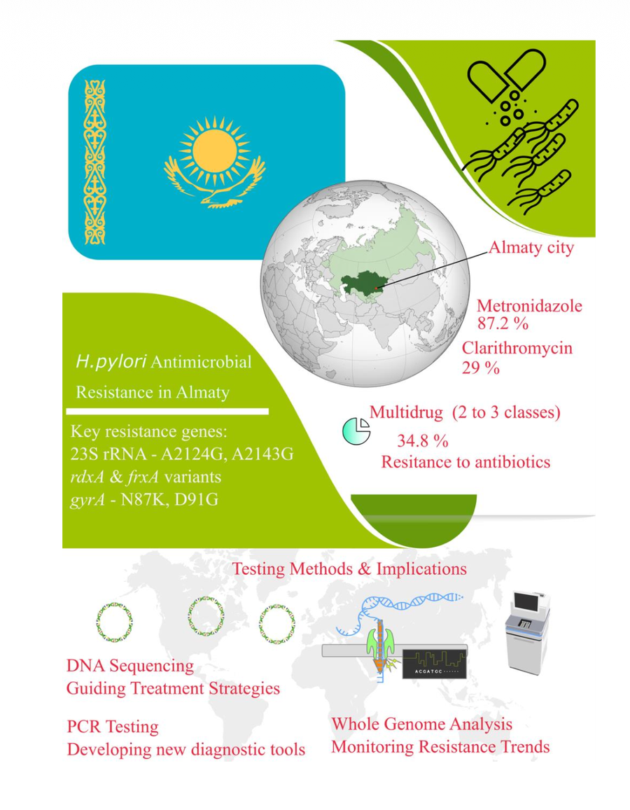
Our single centre culture based study of H. pylori in Kazakhstan, with a regional meta-analysis of prevalence and antibiotic resistance, links the laboratory bench to first line treatment choices in the clinic.
Method
Computation in service of the slide
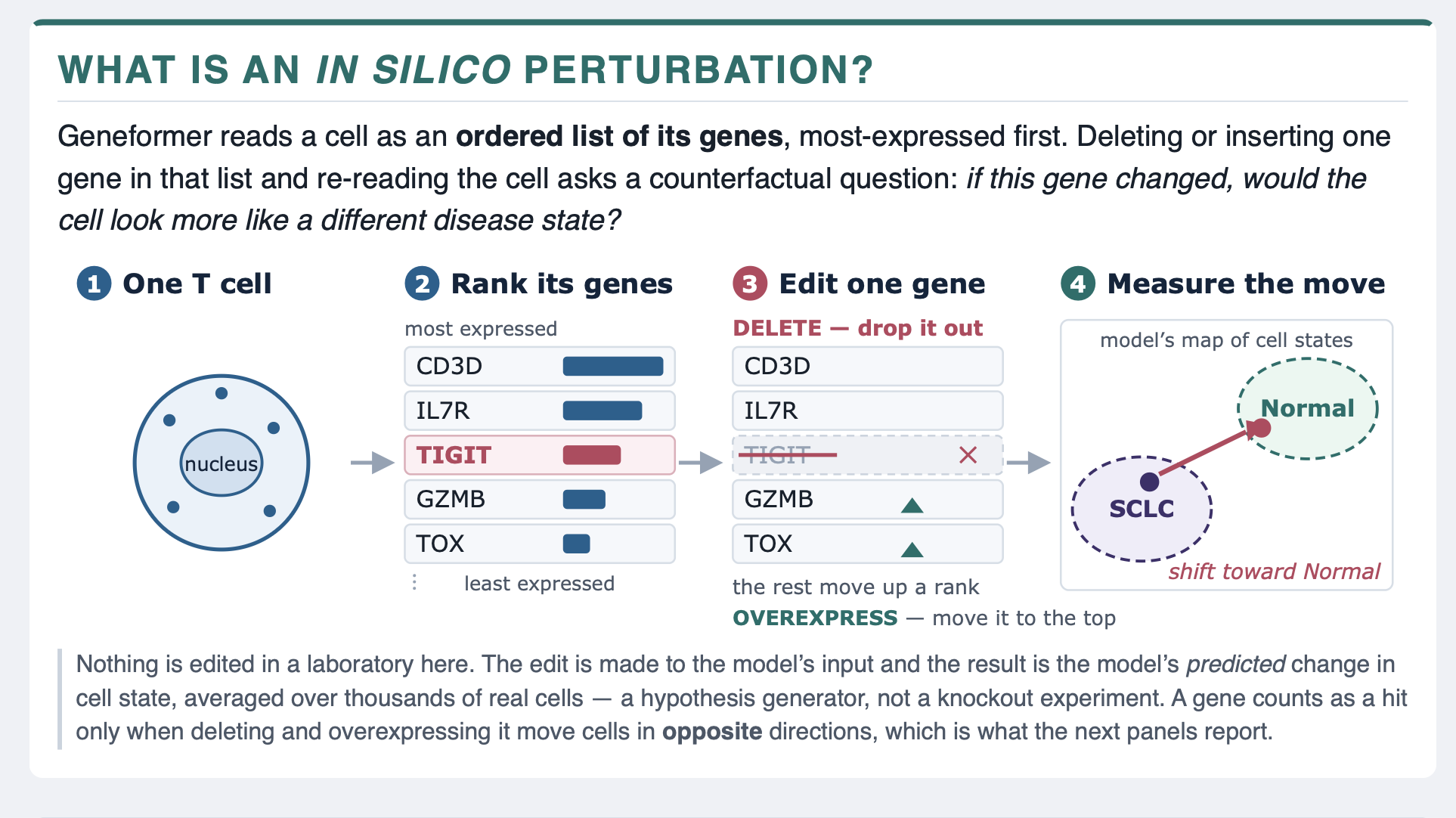
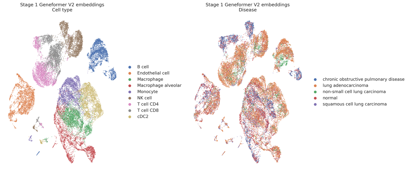
My PhD in genetics and my current work with Geneformer give me tools that most diagnostic teams do not have yet. I use them to generate hypotheses about cell states, then hold those hypotheses to the standard of tissue evidence. A prediction is not a diagnosis.
Immunopathology
Expression profiling in vasculitis
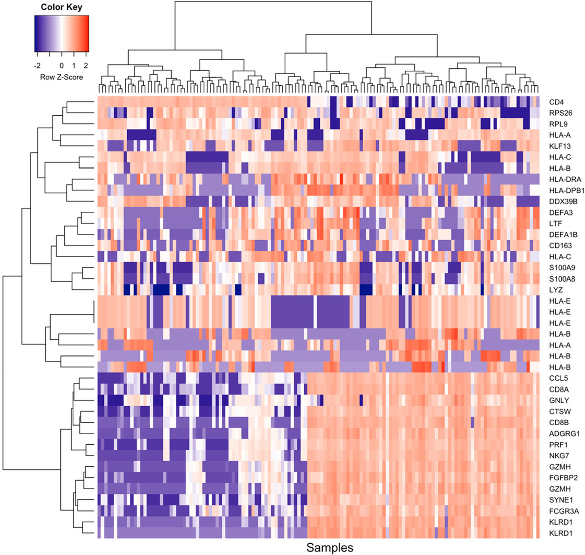
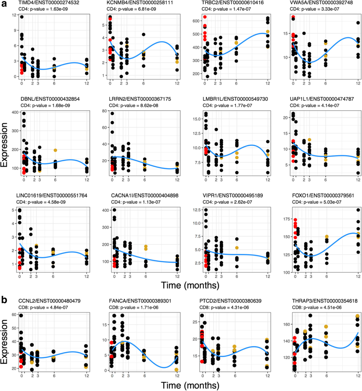
Longitudinal profiling of CD4+ and CD8+ cells in giant cell arteritis, from active disease to remission. My first exposure to reading immune cell states as a marker of tissue disease.
In silico perturbation
Ask the model, then check the tissue
Geneformer predicts how deleting or overexpressing one gene shifts a T cell representation in small cell lung cancer. I check that each donor points the same way, then look for the same signal in the spatial data.
Key figures
Two papers, two ways of reading a signal
Figures
From slide to model to microbe